Search for
DCP-Decarboxy Prothrombin PIVKA-II
Cancer
Report in 72Hrs
At Home
No Fasting Required
Details
Primarily used as a tumor marker for Hepatocellular Carcinoma (HCC)
₹3,990₹4,850
18% OFF
DCP-Decarboxy Prothrombin PIVKA-II Test Information Guide
- Why is it done?
- Detects abnormal prothrombin produced by hepatocellular carcinoma (HCC) and cirrhotic liver tissue
- Screening and surveillance for hepatocellular carcinoma in high-risk patients with chronic liver disease, cirrhosis, and hepatitis B or C
- Early detection of HCC before imaging studies can identify tumors (tumor marker in early stages)
- Monitoring treatment response and disease progression in HCC patients
- Performed at baseline evaluation and during regular surveillance intervals (every 3-6 months) in at-risk populations
- Used in combination with AFP (alpha-fetoprotein) and imaging studies for comprehensive HCC assessment
- Normal Range
- Reference Range: < 40 mAU/mL (milliarbitrary units per milliliter) or < 0.1 ng/mL
- Normal Result: Negative or undetectable DCP levels indicate no evidence of HCC or minimal hepatocellular activity
- Abnormal Result: Elevated levels (≥ 40 mAU/mL) may indicate HCC or advanced cirrhosis
- Borderline Range: 40-400 mAU/mL - warrants careful clinical correlation and possible repeat testing
- Significantly Elevated: > 400 mAU/mL - strongly suggestive of HCC
- Units of Measurement: mAU/mL (milliarbitrary units per milliliter) or ng/mL (nanograms per milliliter)
- Interpretation
- Negative/Normal Results (< 40 mAU/mL): No detectable DCP; unlikely to have HCC at present, though does not completely exclude early disease. May represent normal liver function or benign liver conditions.
- Mildly Elevated (40-100 mAU/mL): May indicate early HCC, advanced cirrhosis, or chronic hepatitis. Requires clinical correlation with imaging and other markers (AFP, ultrasound, CT/MRI).
- Moderately Elevated (100-400 mAU/mL): Moderately suggestive of HCC; strongly warrants confirmatory imaging and detailed evaluation. May also be seen in cirrhosis without HCC.
- Highly Elevated (> 400 mAU/mL): Highly suggestive of HCC; strongly indicative of advanced disease with significant hepatocellular carcinoma burden
- Progressive Elevation: Rising DCP levels on serial testing indicate disease progression or inadequate treatment response
- Declining Levels: Decreasing DCP after treatment may indicate good treatment response, though persistent elevation suggests residual disease
- Factors Affecting Results: Vitamin K deficiency (may falsely elevate), liver function status, extent of cirrhosis, concurrent HBV/HCV infection, chemotherapy, and ablation procedures
- Clinical Significance: DCP has superior specificity for HCC compared to AFP alone, particularly valuable in detecting early-stage HCC. Serial monitoring provides prognostic information and treatment response assessment.
- Associated Organs
- Primary Organ: Liver - DCP is produced by malignant and cirrhotic hepatocytes
- Associated Conditions: Hepatocellular carcinoma (HCC), cirrhosis, chronic hepatitis B, chronic hepatitis C, alcoholic liver disease, non-alcoholic fatty liver disease (NAFLD), hepatic fibrosis, and hepatic inflammation
- Diseases Detected/Monitored: Primary hepatocellular carcinoma, advanced cirrhosis with malignant transformation, recurrent HCC, metastatic disease to liver
- Related Organ Complications: Portal hypertension, esophageal varices, ascites, hepatic encephalopathy, coagulopathy, and hepatorenal syndrome in advanced HCC/cirrhosis
- Metastatic Complications: HCC can metastasize to lungs, bones, brain, and distant lymph nodes; elevated DCP may correlate with metastatic burden
- Follow-up Tests
- Complementary Biomarkers: Alpha-fetoprotein (AFP), AFP-L3 isoform, GP73 (Golgi protein 73), and other serum markers for comprehensive HCC assessment
- Imaging Studies: Ultrasound (US), contrast-enhanced CT (computed tomography), dynamic contrast-enhanced MRI (magnetic resonance imaging) to visualize and characterize liver lesions
- Liver Function Tests: AST, ALT, alkaline phosphatase, bilirubin, albumin, and PT/INR to assess hepatic synthetic function and disease severity
- Viral Serology: Hepatitis B surface antigen (HBsAg), Hepatitis B core antibody (anti-HBc), Hepatitis C antibody (anti-HCV), viral load testing if positive
- Fibrosis Assessment: FIB-4 index, APRI score, elastography (transient or shear wave), or liver biopsy to assess cirrhosis severity
- Biopsy: Liver biopsy if imaging findings are inconclusive and tissue diagnosis is needed to confirm HCC
- Staging Studies: Chest CT, bone scan, or PET-CT for metastatic disease staging in confirmed HCC cases
- Monitoring Frequency: For cirrhotic patients without HCC: every 3-6 months; For HCC patients: every 1-3 months depending on treatment phase and risk; Post-treatment surveillance: as per institutional protocol (typically every 3-4 months for 2 years, then every 4-6 months)
- Related Tumor Markers: Combined score systems (GALAD score, HCC-TORE) using DCP with other markers for improved diagnostic accuracy
- Fasting Required?
- Fasting Required: No - Fasting is NOT required for this test. The test can be performed at any time of day without food or fluid restrictions.
- Specimen Collection: 5-10 mL of venous blood collected in a serum separator tube (SST) or EDTA tube depending on laboratory protocol
- Medications to Avoid: No specific medication restrictions; however, patients on anticoagulants (warfarin, DOACs) should inform the phlebotomist to prevent hematoma formation
- Patient Preparation: Patient should be well-hydrated; sit comfortably for 5 minutes prior to blood draw; inform healthcare provider of any active bleeding disorders or current anticoagulation therapy
- Specimen Handling: Blood sample should be allowed to clot at room temperature for 30 minutes, centrifuged at 1200-1500 g for 10 minutes, and serum stored at 2-8°C if testing is delayed
- Timing Considerations: Test can be performed at any time; consistency in timing (similar time of day) may be preferred for serial monitoring to minimize diurnal variation effects
- Laboratory Turn-Around Time: Typically 2-7 business days; results may be available faster at specialized hepatology centers with in-house testing capabilities
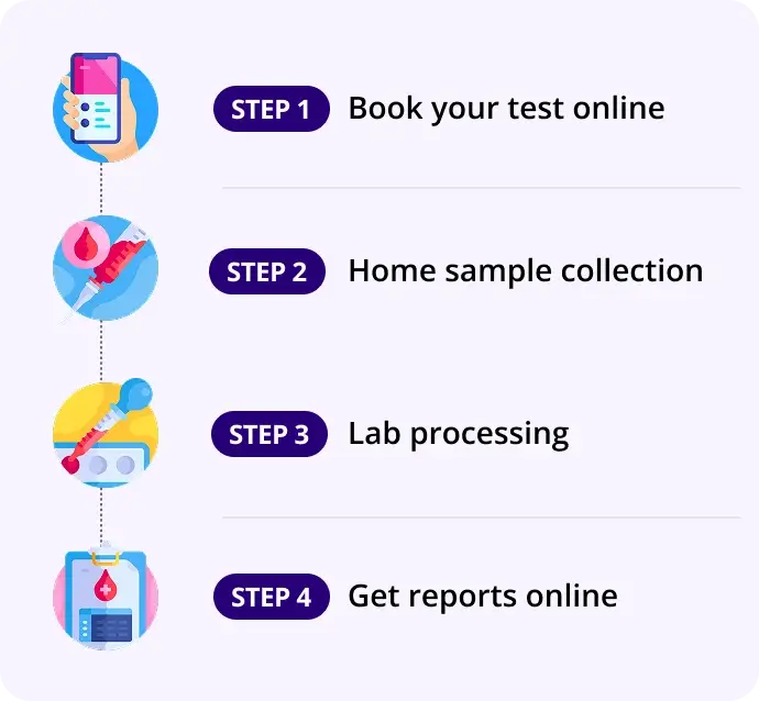
How our test process works!

