Search for
Endometrium biopsy - Medium 1-3 c
Biopsy
Report in 288Hrs
At Home
No Fasting Required
Details
It's commonly performed to investigate abnormal uterine bleeding, infertility, or suspected endometrial pathology (like hyperplasia or cancer).
₹399₹1,000
60% OFF
Endometrium Biopsy - Medium 1-3 c
- Why is it done?
- Test Purpose: Direct tissue examination of the endometrium (uterine lining) to detect cellular abnormalities, malignancy, infections, and structural changes
- Primary Indications: Investigation of abnormal uterine bleeding (AUB), abnormal Pap smear results, detection of endometrial cancer or precancerous lesions, assessment of chronic endometritis, evaluation of infertility, and monitoring of hormone replacement therapy effects
- Typical Timing: Usually performed in premenopausal women during the luteal phase (after ovulation); in postmenopausal women any time during the cycle. Often performed during office gynecologic examination or in conjunction with hysteroscopy
- Medium 1-3 c Specification: Refers to small tissue sample (1-3 cubic centimeters) obtained via endometrial biopsy catheter, providing adequate tissue for histopathological analysis while minimizing patient discomfort
- Normal Range
- Normal Findings: Benign endometrial tissue consistent with proliferative phase (premenopausal) or atrophic endometrium (postmenopausal), without evidence of malignancy, hyperplasia, infection, or significant inflammation
- Reference Range Categories: No specific numerical values; results are qualitative and based on histological examination
- Negative Result Interpretation: No malignancy, no significant hyperplasia, no acute infection, normal benign endometrial tissue appropriate for age and cycle phase
- Positive Result Interpretation: Detection of endometrial cancer, precancerous lesions, significant hyperplasia, chronic endometritis, tuberculosis, or other significant pathology
- Specimen Adequacy: Tissue sample should be 1-3 mm in size with adequate endometrial tissue for histological assessment; samples must be properly fixed and processed for accurate diagnosis
- Interpretation
- Benign Findings: Normal endometrium, inactive/atrophic endometrium, benign polyps, submucosal fibroids, or normal secretory changes indicate no malignancy present
- Hyperplasia Classification: Simple hyperplasia without atypia (low malignant potential), complex hyperplasia without atypia, atypical hyperplasia (increased malignant risk), each requiring different management strategies
- Malignant Findings: Endometrial adenocarcinoma (type I or type II), squamous cell carcinoma, sarcomas, or other malignancies; findings require immediate specialist consultation and treatment planning
- Infectious Processes: Chronic endometritis, tuberculosis, fungal infections, or viral infections may be identified; requires appropriate antimicrobial or antifungal therapy
- Factors Affecting Interpretation: Menstrual cycle phase, hormone therapy use, presence of intrauterine device (IUD), recent curettage, previous radiation, specimen adequacy, and technical processing quality
- Clinical Significance: Results guide treatment decisions, determine need for hysterectomy, guide hormone therapy adjustments, monitor response to previous treatments, and establish prognosis for endometrial pathology
- Inconclusive Results: Inadequate specimen, significant crush artifact, or unclear pathology may necessitate repeat biopsy or alternative imaging studies such as hysteroscopy or curettage
- Associated Organs
- Primary Organ System: Female reproductive system, specifically the uterus and endometrium (inner mucosal lining of the uterine cavity)
- Associated Conditions - Benign: Endometrial polyps, uterine fibroids (leiomyomas), adenomyosis, chronic endometritis, and hormone-related endometrial changes
- Associated Conditions - Malignant: Endometrial cancer (adenocarcinoma being most common), endometrial sarcoma, uterine papillary serous carcinoma (UPSC), clear cell carcinoma, and carcinosarcoma
- Diseases Diagnosed/Monitored: Abnormal uterine bleeding, endometrial hyperplasia, endometrial cancer, polycystic ovary syndrome (PCOS)-related changes, recurrent pregnancy loss, infertility, and hormone-dependent conditions
- Potential Complications from Abnormal Results: Metastatic spread from malignancy, sepsis from untreated infection, infertility from chronic inflammation, recurrent hemorrhage, uterine perforation (during biopsy procedure), and psychological morbidity from cancer diagnosis
- Risk Factors for Abnormal Results: Obesity, diabetes, unopposed estrogen exposure, tamoxifen use, hypertension, PCOS, Lynch syndrome, advanced age, and nulliparity (for malignancy risk)
- Follow-up Tests
- Based on Benign Results: Usually no additional testing required; routine gynecologic surveillance; may consider transvaginal ultrasound if abnormal bleeding persists
- Based on Hyperplasia: Progestin therapy initiation; repeat endometrial biopsy in 3-6 months to assess response; imaging studies if structural abnormalities suspected; gynecologic oncology referral for atypical hyperplasia
- Based on Malignancy: Urgent gynecologic oncology referral; staging computed tomography (CT) or magnetic resonance imaging (MRI); myometrial invasion assessment; lymph node evaluation; tumor marker analysis (CA-125); genetic testing if Lynch syndrome suspected
- Based on Infection: Culture and sensitivity testing if available; targeted antibiotic or antifungal therapy; follow-up biopsy after treatment completion; assessment for underlying causes (IUD-related, tuberculosis transmission)
- Complementary Tests: Transvaginal ultrasound, saline infusion sonography (SIS), hysteroscopy, dilatation and curettage (D&C), endometrial receptivity array (ERA) testing for fertility assessment
- Monitoring Frequency: Benign findings: annual gynecologic exam; Hyperplasia: repeat biopsy 3-6 months post-treatment; Malignancy: oncology-directed surveillance per treatment protocol (typically every 3 months initially)
- Inadequate Specimen: Repeat endometrial biopsy, hysteroscopic-guided biopsy, or dilation and curettage for larger tissue sample and improved diagnostic yield
- Fasting Required?
- Fasting Status: No - fasting is not required for endometrial biopsy
- Pre-Procedure Preparation: Normal diet and fluids permitted; schedule procedure in follicular phase (first 10 days of cycle) when possible; empty bladder and bowels before procedure; wear comfortable, easily removable clothing
- Medications to Continue: Most routine medications may be continued; anticoagulants and antiplatelet agents (aspirin, warfarin, clopidogrel) should be discussed with provider regarding continuation
- Medications to Avoid: Significant anticoagulation therapy may need adjustment; NSAIDs if increased bleeding risk; heavy aspirin use may be discontinued 3-5 days prior
- Pain Management: Ibuprofen 600-800 mg or acetaminophen 500-650 mg may be taken 30-60 minutes before procedure; local anesthetic may be offered during procedure
- Infection Prevention: Prophylactic antibiotics generally not required; may be considered for high-risk patients (immunocompromised, prosthetic heart valve, recent endocarditis)
- Post-Procedure Instructions: Avoid sexual intercourse for 24-48 hours; avoid tampons and douching for several days; normal activities usually permitted same day; some cramping and light spotting expected for 1-2 days
- Pregnancy Considerations: Biopsy should not be performed if pregnancy is suspected; pregnancy test may be recommended prior if cycle is irregular or pregnancy possible
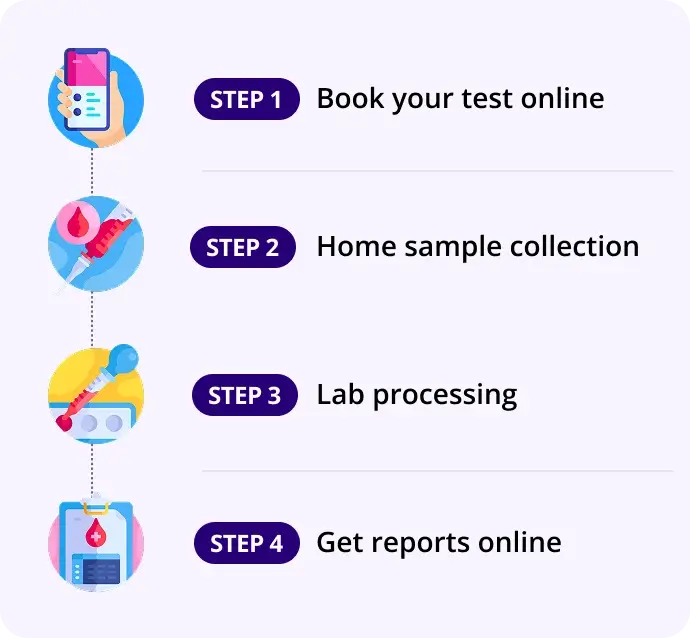
How our test process works!

