Search for
Gall bladder - Large Biopsy 3-6 cm
Biopsy
Report in 288Hrs
At Home
Fasting Required
Details
Confirm or rule out malignancy (especially gall bladder carcinoma)
₹699₹1,500
53% OFF
Gallbladder - Large Biopsy 3-6 cm
- Why is it done?
- Diagnostic evaluation of suspicious gallbladder lesions or masses identified on imaging studies
- Assessment of gallbladder wall thickening or nodular lesions to determine benign versus malignant pathology
- Detection and characterization of adenocarcinoma, cholangiocarcinoma, or other malignant tumors
- Investigation of gallbladder polyps >6 mm or those showing growth on serial imaging
- Histopathological diagnosis when clinical and radiological findings are inconclusive
- Typically performed when ultrasound, CT, or MRI imaging reveals lesions measuring 3-6 cm requiring tissue confirmation
- Assessment of gallbladder inflammation or suspected cholecystitis when imaging is equivocal
- Normal Range
- Normal Result: Benign gallbladder tissue or absence of malignancy
- Specimen Quality: Adequate tissue sample of 3-6 cm with proper fixation and preservation
- Negative for Malignancy: Histological examination reveals no cancerous cells or dysplasia
- Benign Findings: Cholesterol polyps, adenomyomatosis, or chronic cholecystitis without atypia
- Normal Gallbladder Wall: Histologically intact epithelium without inflammation or proliferative lesions
- Interpretation
- Malignant Findings: Presence of adenocarcinoma, squamous cell carcinoma, cholangiocarcinoma, or other malignant histology indicates gallbladder cancer requiring immediate treatment planning and oncology consultation
- Dysplasia: Low-grade or high-grade dysplasia indicates precancerous changes; high-grade dysplasia warrants cholecystectomy to prevent progression to invasive cancer
- Benign Polyps: Cholesterol polyps or inflammatory polyps without dysplasia typically have excellent prognosis; conservative management or surveillance ultrasound may be appropriate
- Adenomyomatosis: Benign condition with muscular hyperplasia and glandular proliferation; generally does not require treatment unless symptomatic
- Chronic Cholecystitis: Inflammatory changes without dysplasia indicate chronic inflammation, often associated with gallstones; management depends on clinical symptoms
- Inconclusive/Non-Diagnostic: Insufficient tissue or inadequate sampling may require repeat biopsy or alternative imaging follow-up
- Factors Affecting Results: Biopsy location within lesion, tissue preservation quality, specimen adequacy, and pathologist expertise may influence accuracy; multiple biopsies improve diagnostic yield
- Associated Organs
- Primary Organ: Gallbladder (biliary system)
- Associated Biliary Structures: Common bile duct, hepatic ducts, and ampulla of Vater; tumors may involve or extend to these structures
- Related Organs: Liver, pancreas, and duodenum; gallbladder cancer may metastasize to these organs or involve them directly
- Conditions Diagnosed: Gallbladder adenocarcinoma, cholangiocarcinoma, intrahepatic cholangiocarcinoma, gallbladder metastases, benign polyps, adenomyomatosis, and chronic cholecystitis
- Risk Factors for Gallbladder Cancer: Cholelithiasis (gallstones), primary sclerosing cholangitis, chronic hepatitis, cirrhosis, and choledochal cysts increase malignancy risk
- Potential Complications: Biliary obstruction causing jaundice and cholangitis, hepatic dysfunction, pancreatitis, and peritoneal involvement in advanced malignancy
- Follow-up Tests
- If Malignancy Confirmed: CT abdomen/pelvis with IV contrast for staging, MRI/MRCP for biliary tract involvement, and PET-CT for metastatic disease assessment
- Laboratory Tests: Liver function tests (bilirubin, alkaline phosphatase, transaminases), tumor markers (CA 19-9, CEA), and complete metabolic panel
- If Dysplasia Present: Cholecystectomy with histopathological examination of entire specimen; high-grade dysplasia is indication for surgical resection
- If Benign Findings: Surveillance ultrasound at 6 months then annually for polyps <10 mm; cholecystectomy considered if symptoms develop or polyp growth occurs
- Immunohistochemistry: May be performed on biopsy samples to characterize tumor phenotype and determine immunotherapy eligibility
- Molecular Testing: Genetic mutations (KRAS, TP53, CDKN2A) may be analyzed for prognostic information and targeted therapy options
- Monitoring Schedule: Post-operative imaging and serum markers every 3-4 months for first year if cancer diagnosed; surveillance imaging every 6-12 months for benign conditions
- Fasting Required?
- Fasting Status: YES - Fasting is required
- Fasting Duration: Minimum 6-8 hours of NPO (nothing by mouth) status before procedure; typically overnight fasting recommended
- Special Instructions: No food, beverages, or chewing gum after midnight before morning procedure; water may be permitted up to 2-3 hours before procedure (confirm with facility)
- Medications: Anticoagulants (warfarin, direct oral anticoagulants) and antiplatelet agents (aspirin, clopidogrel) should be discontinued 5-7 days before procedure; consult with physician regarding specific medications
- Pre-Procedure Preparation: Informed consent required; review risks (bleeding, infection, bile duct injury, pancreatitis); arrange transportation as sedation may be used; baseline coagulation studies (INR, PT/PTT) typically ordered
- Post-Procedure Instructions: Resume normal diet and medications after procedure per provider instructions; monitor for fever, severe abdominal pain, or jaundice; rest for 24 hours; avoid strenuous activity for several days
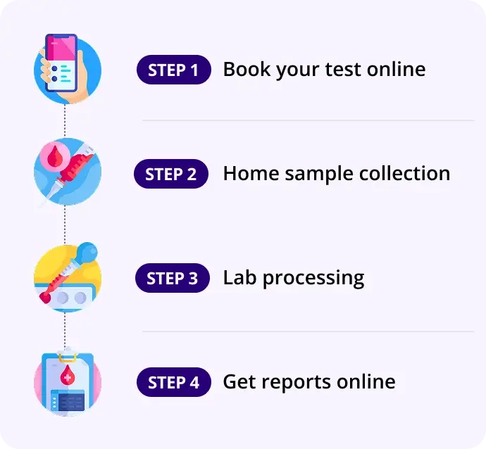
How our test process works!

