Blog Summary
Metabolic dysfunction is the silent engine driving three of today's most dangerous chronic diseases: type 2 diabetes, cardiovascular disease, and non-alcoholic fatty liver disease. This thorough, science-based guide covers the clinical mechanisms by which poor metabolism contributes to each condition, how these diseases feed each other over time, and evidence-based prevention and management strategies drawn from peer-reviewed research by the ADA, AHA, NHLBI, and the Diabetes Prevention Program.
1. Introduction: The Metabolic Crisis Reshaping Global Health
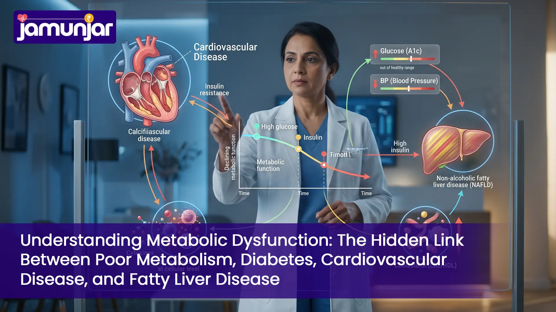
Metabolic health is one of the most pressing challenges confronting modern medicine. Metabolic syndrome — a cluster of cardiometabolic risk factors occurring together — affects an estimated 80 million Americans alone. Yet despite its prevalence, many people do not realise that poor metabolism is the underlying mechanism linking three of the most common chronic diseases: type 2 diabetes mellitus (T2DM), cardiovascular disease (CVD), and non-alcoholic fatty liver disease (NAFLD).
Understanding the complex pathophysiological relationships between metabolic dysfunction and these chronic comorbidities is essential for both prevention and early intervention. This guide covers the mechanistic links, diagnostic criteria, and evidence-based treatment approaches to support informed decision-making for both clinicians and patients. For individuals in metro cities with multiple risk factors, services such as physiotherapy at home in Mumbai can be highly beneficial in establishing the physical activity patterns that metabolic health depends on.
2. What Is Metabolic Dysfunction? A Clinical Definition and Mechanistic Overview
2.1 Defining Metabolic Dysfunction at the Cellular Level
Metabolic dysfunction encompasses impaired cellular energy metabolism with decreased insulin sensitivity, aberrant glucose homeostasis, and dysregulated lipid metabolism. At the heart of this impairment is a failure of mitochondrial functioning and insulin signalling cascades.
The central mechanism is insulin resistance — a state in which cells do not respond properly to insulin signals. When this occurs across muscle, liver, and adipose tissue, several pathogenic cascades are initiated:
- Impaired peripheral glucose absorption with compensatory increase in insulin production by the pancreas (hyperinsulinemia)
- Elevated blood glucose with increased hepatic glucose synthesis
- Increased lipolysis in adipose tissue, raising circulating free fatty acid levels
- Elevated pro-inflammatory cytokines including IL-6 and TNF-α
2.2 Molecular and Biochemical Markers of Metabolic Dysfunction
Key clinical indicators of metabolic dysfunction include:
- Fasting insulin and HOMA-IR (Homeostasis Model Assessment for Insulin Resistance)
- Elevated triglycerides and low HDL cholesterol
- Elevated blood pressure and fasting glucose
- Raised inflammatory markers including high-sensitivity CRP, fibrinogen, and homocysteine
3. Metabolic Syndrome: Understanding the Gateway to Disease
3.1 Diagnostic Criteria for Metabolic Syndrome
Metabolic syndrome is a clustering of metabolic disorders that significantly increases the risk of T2DM and CVD. Per the International Diabetes Federation (IDF) and NHLBI, the diagnosis requires central obesity plus any two of the following four additional findings:
- Central Obesity: Waist circumference >40 inches (men) or >35 inches (women) — reflects visceral fat accumulation and impaired adipose tissue function
- Elevated Triglycerides: ≥150 mg/dL or on lipid-lowering medication — reflects hepatic lipid overflow and impaired lipolysis
- Reduced HDL Cholesterol: <40 mg/dL (men) or <50 mg/dL (women) — loss of the anti-atherogenic lipoprotein fraction
- Elevated Blood Pressure: ≥130/85 mmHg or on antihypertensive medication — reflects sympathetic nervous system activation
- Elevated Fasting Glucose: ≥100 mg/dL or existing T2DM diagnosis — indicates beta-cell compensation for insulin resistance
3.2 Epidemiological Impact
Approximately 34% of US adults have metabolic syndrome. Research published in JAMA has shown that individuals with metabolic syndrome face a 3.7 times higher risk of myocardial infarction and a 2.6 times higher cardiovascular mortality risk compared to those without the condition.
4. How Poor Metabolism Leads to Type 2 Diabetes
4.1 The Pathophysiology of Insulin Resistance and Beta-Cell Dysfunction
The development from metabolic dysfunction to type 2 diabetes is characterised by two fundamental pathogenic processes:
Insulin Resistance: The primary defect occurs in peripheral tissue — skeletal muscle, liver, and adipose tissue. Normal insulin signalling involves a cascade through phosphatidylinositol 3-kinase (PI3K) and protein kinase B (Akt), culminating in glucose transporter 4 (GLUT4) translocation and glucose uptake. In metabolic dysfunction, this pathway becomes dysfunctional, reducing glucose clearance and driving compensatory hyperinsulinemia.
Beta-Cell Exhaustion: Pancreatic beta cells initially compensate by increasing insulin output. Over time, persistent hyperglycaemia and oxidative stress cause beta-cell apoptosis and functional decline. Studies using isolated islets have demonstrated that chronic hyperglycaemia reduces mitochondrial ATP production and impairs glucose-stimulated insulin secretion. After 10 to 15 years of insulin resistance, beta-cell compensation eventually fails, resulting in overt hyperglycaemia and a clinical diagnosis of diabetes.
4.2 Contributing Molecular Mechanisms
- Free Fatty Acid Toxicity (Lipotoxicity): Elevated circulating free fatty acids impair insulin signalling via serine/threonine kinase pathways, including c-Jun N-terminal kinase (JNK), leading to insulin receptor substrate 1 (IRS-1) degradation
- Endoplasmic Reticulum (ER) Stress: Chronic dietary excess induces unfolded protein response (UPR) pathways, increases JNK phosphorylation and NF-κB activation, and amplifies insulin resistance
- Mitochondrial Dysfunction: Decreased oxidative phosphorylation capacity leads to impaired ATP generation, reduced glucose-stimulated insulin secretion, and metabolic inflexibility
- Low-Grade Chronic Inflammation: Elevated TNF-α, IL-6, and CRP limit IRS-1 tyrosine phosphorylation and GLUT4 translocation
4.3 Timeline and Progression
The progression from metabolic dysfunction to T2DM is a continuum:
- Years 1–5: Insulin resistance develops; fasting glucose and triglycerides begin rising; fasting insulin above 12 mU/L
- Years 5–10: Postprandial hyperglycaemia emerges; HbA1c rises to 5.7–6.4%; prediabetes is diagnosed
- Years 10–15: Beta-cell compensation fails; fasting glucose reaches ≥126 mg/dL; type 2 diabetes is diagnosed
5. The Cardiovascular Connection: Metabolic Dysfunction and Heart Disease
5.1 Metabolic Dysfunction as an Independent Cardiovascular Risk Factor
The link between poor metabolism and cardiovascular disease is well established in the epidemiological literature. The Framingham Heart Study and the Nurses' Health Study both demonstrated that metabolic dysfunction is an independent predictor of cardiovascular events, even in the absence of overt diabetes or hypertension.
5.2 Mechanisms Linking Metabolic Dysfunction to Atherosclerosis
Dyslipidaemia and LDL Particle Composition: Insulin resistance increases hepatic VLDL synthesis and reduces lipoprotein lipase activity, resulting in elevated triglycerides, low HDL cholesterol, and a shift toward small, dense LDL particles — the most atherogenic LDL phenotype. These particles are more likely to penetrate the arterial intima and are more susceptible to oxidation.
Endothelial Dysfunction: Hyperinsulinemia and elevated glucose reduce nitric oxide (NO) bioavailability, impairing endothelial-dependent vasodilation. Insulin resistance also increases arginase activity, competing with endothelial nitric oxide synthase (eNOS) for L-arginine and further reducing NO production. Decreased HDL activation of eNOS and increased oxidative stress via NADPH oxidase activation further compound this dysfunction.
Vascular Inflammation and Atherosclerotic Lesion Development: Metabolic dysfunction amplifies monocyte recruitment into the arterial wall through upregulated expression of adhesion molecules (ICAM-1, VCAM-1). Once in the intima, monocytes differentiate into macrophages that ingest oxidised LDL particles, forming foam cells — the hallmark of early atherosclerotic lesions. Pro-inflammatory cytokines including TNF-α and IL-6 sustain this process.
Thrombotic Abnormalities: Metabolic dysfunction raises prothrombotic factors including fibrinogen and impairs tissue plasminogen activator (tPA) activity, while promoting platelet dysfunction. These haemostatic changes increase the risk of thrombotic events at vulnerable atherosclerotic plaques.
5.3 Clinical Cardiovascular Outcomes
Research published in Circulation has shown that people with metabolic syndrome are 2.88 times more likely to develop coronary artery disease and 2.43 times more likely to suffer a stroke. In patients with acute coronary syndrome, the presence of metabolic syndrome is associated with more severe coronary disease and worse long-term outcomes.
6. Non-Alcoholic Fatty Liver Disease (NAFLD): The Silent Metabolic Consequence
6.1 Epidemiology and Pathogenesis
NAFLD — increasingly referred to as metabolic-associated fatty liver disease (MAFLD) — encompasses a spectrum of hepatic lipid accumulation ranging from simple steatosis to non-alcoholic steatohepatitis (NASH), with the potential to progress to cirrhosis and hepatocellular carcinoma (HCC). NAFLD affects an estimated 25 to 30% of the global population, rising to over 75% in communities with high rates of obesity and T2DM.
6.2 The "Two-Hit" Model of NAFLD Pathogenesis
First Hit — Hepatic Steatosis: Insulin resistance in the liver impairs regulation of acetyl-CoA carboxylase (ACC) activity and stimulates de novo lipogenesis (DNL). Combined with decreased mitochondrial fatty acid oxidation and increased hepatic uptake of free fatty acids, this results in excess triglyceride accumulation in hepatocytes.
Second Hit — Hepatic Inflammation and Oxidative Stress: Excess hepatic lipids undergo lipid peroxidation, generating reactive species that cause mitochondrial dysfunction. Lipopolysaccharides (LPS) from dysbiotic gut microbiota further aggravate hepatic inflammation via toll-like receptor 4 (TLR4) activation. The resulting oxidative stress activates hepatic stellate cells (HSCs), leading to collagen deposition and fibrosis.
Recent research supports a more comprehensive multihit model that also incorporates metabolic endotoxemia, adipose tissue inflammation, genetic and epigenetic variables, and gut microbiome alterations.
6.3 Diagnostic Considerations
Liver biopsy remains the gold standard for differentiating simple steatosis from NASH, but non-invasive tools are increasingly used in clinical practice:
- FIB-4 Index: (Age × AST) / (Platelet count × √ALT) — predicts advanced fibrosis
- APRI Score: (AST/upper normal limit) / Platelet count × 100
- Transient Elastography (FibroScan): Liver stiffness measurement with strong correlation to fibrosis stage
- Magnetic Resonance Elastography (MRE): High specificity for fibrosis detection and staging
7. The Bidirectional Relationship: How These Diseases Amplify Each Other
7.1 Diabetes Exacerbating Cardiovascular Risk
Cardiovascular risk rises substantially with the onset of type 2 diabetes. Chronic hyperglycaemia directly injures the endothelium via the polyol pathway, advanced glycation end products (AGEs), and protein kinase C (PKC) activation. Diabetes is also an independent risk factor for atrial fibrillation, accounting for approximately 10% of AF occurrences. The combination of metabolic dysfunction, diabetes, and cardiac arrhythmia represents a particularly high-risk clinical phenotype.
7.2 Fatty Liver Disease Accelerating Metabolic Dysfunction and Diabetes Progression
NAFLD aggravates metabolic dysfunction through several bidirectional pathways:
- Worsening hepatic insulin resistance: Accumulated hepatic lipids and inflammation impair insulin receptor phosphorylation and downstream signalling
- Increased hepatic glucose output: Hyperinsulinemia fails to suppress hepatic gluconeogenesis, worsening hyperglycaemia
- Dysbiosis-related metabolic endotoxemia: NAFLD-associated gut dysbiosis increases intestinal permeability, amplifying LPS absorption and TLR4 activation
8. Risk Factors That Accelerate Metabolic Decline
8.1 Modifiable Risk Factors
- Physical Inactivity: Sedentary behaviour impairs skeletal muscle insulin sensitivity by reducing GLUT4 translocation and glucose oxidation capacity. Regular physical activity improves insulin sensitivity independently of weight change.
- High-Calorie Ultra-Processed Diet: Refined carbohydrates and added sugars drive postprandial hyperglycaemia and beta-cell stress. High saturated fat intake promotes hepatic steatosis and systemic inflammation.
- Sleep Dysfunction: Chronic sleep deprivation (under 7 hours nightly) impairs glucose tolerance, elevates cortisol and ghrelin, and promotes visceral adiposity.
- Chronic Psychological Stress: Sustained stress raises cortisol, promoting insulin resistance, visceral fat deposition, and low-grade inflammation.
- Tobacco Use: Smoking reduces pancreatic beta-cell function and increases insulin resistance and atherosclerosis through oxidative stress mechanisms.
- Excessive Alcohol Use: Heavy alcohol intake contributes to hepatic steatosis and systemic inflammation.
8.2 Non-Modifiable Risk Factors
- Age: Insulin sensitivity naturally declines with age, in part due to reduced physical activity and sarcopenia.
- Ethnicity: South Asian, Hispanic, and African American populations show higher prevalence of metabolic syndrome, partly due to genetic predisposition to insulin resistance.
- Family History: Twin studies demonstrate 50 to 70% heritability of insulin resistance and metabolic syndrome susceptibility.
- Maternal Factors: Prenatal programming, intrauterine undernutrition, and maternal obesity increase the risk of metabolic disorders in offspring through epigenetic alterations.
9. Evidence-Based Diagnostic Approaches
9.1 Screening Recommendations
The ADA and AHA recommend screening for metabolic dysfunction in:
- All adults aged 45 or older, or those under 45 with BMI ≥25 and additional risk factors
- Individuals with hypertension, dyslipidaemia, or polycystic ovarian syndrome (PCOS)
- First-degree relatives of people with type 2 diabetes
9.2 Core Diagnostic Tests
- Fasting Glucose and HbA1c: Evaluate glucose management and prediabetes/diabetes status
- Fasting Lipid Panel: Total cholesterol, LDL, HDL, and triglycerides
- Blood Pressure Measurement: Both in-office and ambulatory measurements
- Body Composition Assessment: BMI, waist circumference, and ideally bioelectrical impedance analysis (BIA) or DEXA for visceral fat quantification
- Liver Function Tests: ALT, AST, GGT, and hepatitis B and C serology
- Inflammatory Markers: High-sensitivity CRP for cardiovascular risk stratification
9.3 Advanced Diagnostic Modalities
- Oral Glucose Tolerance Test (OGTT): Gold standard for detecting impaired glucose tolerance and early diabetes
- Insulin and C-Peptide Levels: Assess beta-cell function and degree of insulin resistance
- Advanced Lipid Testing: LDL particle number, size, and apolipoprotein B (ApoB) measurements
- Coronary Artery Calcium (CAC) Score: Identifies subclinical atherosclerosis in asymptomatic individuals
10. Comprehensive Prevention and Management Strategies
10.1 Lifestyle Modification Interventions
The landmark Diabetes Prevention Program (DPP) demonstrated that intensive lifestyle modification — including modest weight loss of 7% of baseline body weight, 150 minutes of aerobic exercise per week, and dietary change — reduced the risk of diabetes by 58%, rising to 71% in those over 65. This remains the gold standard intervention for preventing metabolic dysfunction from progressing.
Nutritional Approaches
- Mediterranean Diet: The most evidence-supported pattern for cardiovascular health; emphasises olive oil, legumes, whole grains, fish, and vegetables
- DASH Diet: Clinically proven to lower blood pressure and improve lipid profiles; recommended by AHA
- Low Glycaemic Index/Load: Reduces postprandial glucose excursions and improves HbA1c
- Protein and Fibre Focus: Improves satiety, supports glucose management, and promotes healthier lipid profiles
Physical Activity Recommendations
- Aerobic Exercise: 150 minutes of moderate-intensity exercise per week improves insulin sensitivity through increased GLUT4 expression and mitochondrial biogenesis
- Resistance Training: 2 to 3 sessions per week preserve lean muscle mass, which is critical for glucose homeostasis and insulin sensitivity
- High-Intensity Interval Training (HIIT): Superior to continuous moderate-intensity exercise for enhancing insulin sensitivity and beta-cell function
Structured movement under professional supervision is especially valuable for those with joint discomfort, deconditioning, or recovery from illness. Physiotherapy at home in Delhi and similar services allow patients to begin and maintain safe, prescribed exercise regimens that directly improve insulin sensitivity and cardiovascular outcomes.
Sleep and Stress Management
- Sleep Hygiene: Targeting 7 to 9 hours of sleep nightly with a consistent sleep-wake schedule
- Stress Reduction: Mindfulness-based stress reduction (MBSR), cognitive behavioural therapy (CBT), and yoga are clinically shown to reduce cortisol and improve metabolic parameters
10.2 Pharmacological Interventions
While lifestyle modification remains the cornerstone, pharmacological treatments play an important role in high-risk individuals or those unable to reach targets through lifestyle changes alone.
For Glycaemic Control
- GLP-1 Receptor Agonists: Reduce cardiovascular events and support weight loss alongside glycaemic management; recommended as first-line in patients with T2DM and established CVD
- SGLT2 Inhibitors: Reduce cardiovascular events and slow diabetic kidney disease progression, with modest weight loss and blood pressure reduction
- Metformin: First-line agent that improves insulin sensitivity and reduces cardiovascular events
For Cardiovascular Risk Reduction
- Statins: Reduce LDL cholesterol by 30 to 50%; indicated for all patients with metabolic syndrome and diabetes
- Antihypertensive Agents: ACE inhibitors and ARBs offer renal and cardiovascular protection
- Antiplatelet Therapy: Aspirin considered for primary prevention in high-risk individuals
For Hepatic Steatosis and NAFLD
- Pioglitazone: Improves hepatic insulin sensitivity and reduces hepatic lipid content; beneficial in NASH
- GLP-1 Receptor Agonists: Growing evidence for use in NASH, with weight loss and broader metabolic benefits
11. Frequently Asked Questions
Can Fatty Liver Cause Diabetes?
Yes. There is substantial evidence of a reciprocal relationship. NAFLD induces hepatic and systemic insulin resistance through several mechanisms, including hepatic lipid accumulation, inflammation, and dysbiosis-related metabolic endotoxemia. Mendelian randomisation studies confirm a causal association between liver fat content and incident type 2 diabetes, independent of obesity. Since both conditions share underlying metabolic dysfunction, causality operates in both directions.
What Is the Connection Between Diabetes and Heart Disease?
Several mechanisms drive atherosclerosis in type 2 diabetes: chronic hyperglycaemia directly damages the endothelium; hyperinsulinemia and insulin resistance promote small dense LDL formation and impair HDL function; and inflammatory mediators propagate atherosclerotic plaque development. Additionally, diabetes increases thrombotic risk through platelet dysfunction and elevated fibrinogen. This results in more aggressive and earlier-onset coronary artery disease — up to 50% of patients may experience a myocardial infarction within 10 years of a diabetes diagnosis if left untreated.
Does Fatty Liver Cause Low Metabolism?
NAFLD does not lower basal metabolic rate in the classical sense, but it can significantly impair cellular energy metabolism and contribute to metabolic inflexibility — the inability to switch efficiently between carbohydrate and fat oxidation. In NASH, hepatic mitochondrial dysfunction reduces ATP generation capacity, and accumulating liver lipids interfere with insulin signalling pathways crucial for glucose metabolism. This metabolic inflexibility perpetuates insulin resistance in a self-amplifying cycle and makes weight loss progressively more difficult. Visceral adiposity, which is common in NAFLD, further compounds this metabolic rigidity.
How Does Metabolism Cause Diabetes?
Diabetes is the time-dependent end result of metabolic dysfunction — specifically progressive insulin resistance combined with declining insulin secretion capacity. Initially, increased beta-cell insulin production compensates for peripheral resistance. Over years to decades, chronic hyperglycaemia and oxidative stress cause beta-cell apoptosis and functional decline. When beta-cell function falls below the threshold needed to maintain glucose homeostasis, fasting glucose rises above 126 mg/dL — the diagnostic threshold for type 2 diabetes. The DPP study demonstrated a 58 to 71% risk reduction with lifestyle intervention, confirming that this process is both preventable and modifiable.
If you are beginning a metabolic recovery journey, physiotherapy at home in Hyderabad offers structured physical rehabilitation and guided movement therapy — both clinically proven strategies to improve insulin sensitivity and support long-term metabolic outcomes.
12. Medical References
- Alberti KGMM, Eckel RH, Grundy SM, et al. Harmonizing the Metabolic Syndrome: A Joint Interim Statement of the IDF Task Force on Epidemiology and Prevention. Circulation. 2009;120(16):1640–1645. Available from: https://pubmed.ncbi.nlm.nih.gov/19805654/
- Knowler WC, Barrett-Connor E, Fowler SE, et al. Reduction in the Incidence of Type 2 Diabetes with Lifestyle Intervention or Metformin. New England Journal of Medicine. 2002;346(6):393–403. Available from: https://pubmed.ncbi.nlm.nih.gov/11832527/
- Isomaa B, Almgren P, Tuomi T, et al. Cardiovascular Morbidity and Mortality Associated with the Metabolic Syndrome. Diabetes Care. 2001;24(4):683–689. Available from: https://pubmed.ncbi.nlm.nih.gov/11315831/
- Kahn SE, Zraika S, Utzschneider KM, Hull RL. The Beta Cell Lesion in Type 2 Diabetes: There Has to Be a Primary Functional Abnormality. Diabetologia. 2009;52(6):1003–1012. Available from: https://pubmed.ncbi.nlm.nih.gov/19437789/
- Perseghin G, Ghosh S, Gerow K, Shulman GI. Metabolic Defects in Lean Nondiabetic Offspring of NIDDM Parents. Diabetes. 1997;46(6):1001–1009. Available from: https://pubmed.ncbi.nlm.nih.gov/9166673/
- Kannel WB, McGee DL. Diabetes and Cardiovascular Disease: The Framingham Study. JAMA. 1979;241(19):2035–2038. Available from: https://pubmed.ncbi.nlm.nih.gov/430798/
- Younossi ZM, Koenig AB, Abdelatif D, et al. Global Epidemiology of Nonalcoholic Fatty Liver Disease. Hepatology. 2016;64(1):73–84. Available from: https://pubmed.ncbi.nlm.nih.gov/26926713/
- Tilg H, Adolph TE, Moschen AR. Multiple Parallel Hits Hypothesis in Nonalcoholic Fatty Liver Disease. Journal of Hepatology. 2021;75(3):568–580. Available from: https://pubmed.ncbi.nlm.nih.gov/34048773/
- American Diabetes Association. Standards of Care in Diabetes. Diabetes Care. 2023;46(Suppl 1):S1–S291. Available from: https://pmc.ncbi.nlm.nih.gov/articles/PMC9810475/
- Grundy SM, Stone NJ, Bailey AL, et al. 2018 AHA/ACC Guideline on the Management of Blood Cholesterol. Journal of the American College of Cardiology. 2019;73(25):e285–e350. Available from: https://pubmed.ncbi.nlm.nih.gov/30423393/
Medical Disclaimer
This article is for educational purposes only and does not replace professional medical advice, diagnosis, or treatment. Exercise and dietary changes should be individualised, especially for people with diabetes, cardiovascular disease, joint pain, or limited mobility.